41 parts of the eye without labels
Anatomy of the Eye | Kellogg Eye Center | Michigan Medicine Structure containing muscle and is located behind the iris, which focuses the lens. Cornea The clear front window of the eye which transmits and focuses (i.e., sharpness or clarity) light into the eye. Corrective laser surgery reshapes the cornea, changing the focus. Fovea The center of the macula which provides the sharp vision. Iris Anatomy of the Eye. Learn about the different parts of the eye. The sclera is a membrane of tendon in the eye, also known as the white of the eye. Rugged and robust, the sclera works to protect the inner, more sensitive parts of the eye like the retina and choroid. It is about 0.03 of an inch thick except for where the four "straight" eye muscles append, where the depth is no more than 0.01 of an inch.
Cornea of the Eye - Definition and Detailed Illustration The cornea is the clear front surface of the eye. It lies directly in front of the iris and pupil, and it allows light to enter the eye. Viewed from the front of the eye, the cornea appears slightly wider than it is tall. This is because the sclera (the "white" of the eye) slightly overlaps the top and bottom of the anterior cornea.

Parts of the eye without labels
Learn the Nine Essential Parts of Eyeglasses 1. Rims The rims lend form and character to your eyeglasses—they also provide function by holding the lenses in place. 2. End pieces The end pieces are the small parts on the frame that extend outward and connect the lenses to the temples. 3. Bridge The bridge is the center of the frame that rests on your nose and joins the two rims together. 4. Iris of the Eye: Definition, Anatomy & Common Conditions - Cleveland Clinic The iris is the colored part of your eye. Muscles in your iris control your pupil. ... Some people are born without an iris in one or both of their eyes — a genetic condition called aniridia. Without an iris, your eye would still function, but your vision would be blurry. ... Wear sunglasses with 100% UV protection or a UV400 label anytime ... Label the Eye - The Biology Corner Label the Eye. Shannan Muskopf December 30, 2019. This worksheet shows an image of the eye with structures numbered. Students practice labeling the eye or teachers can print this to use as an assessment. There are two versions on the google doc and pdf file, one where the word bank is included and another with no word bank for differentiation.
Parts of the eye without labels. The Eyes (Human Anatomy): Diagram, Optic Nerve, Iris, Cornea ... - WebMD The front part (what you see in the mirror) includes: Iris: the colored part. Cornea: a clear dome over the iris. Pupil: the black circular opening in the iris that lets light in. Sclera: the ... Structure and Functions of Human Eye with labelled Diagram - BYJUS The internal components of the eye include: Lens Retina Aqueous humour Optic nerve Vitreous humour Test your knowledge on Structure Of Eye Put your understanding of this concept to test by answering a few MCQs. Click 'Start Quiz' to begin! Select the correct answer and click on the "Finish" button Check your score and answers at the end of the quiz Eye Anatomy: A Closer Look At the Parts of the Eye - All About Vision Eye anatomy: A closer look at the parts of the eye. When surveyed about the five senses — sight, hearing, taste, smell and touch — people consistently report that their eyesight is the mode of perception they value (and fear losing) most. Despite this, many people don't have a good understanding of the anatomy of the eye, how vision works ... Your Eyes (for Kids) - Nemours KidsHealth The white part of the eyeball is called the sclera (say: SKLAIR-uh). The sclera is made of a tough material and has the important job of covering most of the eyeball. Think of the sclera as your eyeball's outer coat. Look very closely at the white of the eye, and you'll see lines that look like tiny pink threads.
Blood vessels and nerves of the eye: Anatomy | Kenhub The main blood supply of the eye arises from the ophthalmic artery, which gives off orbital and optical group branches. Innervation of the eyeball and surrounding structures is provided by the optic, oculomotor, trochlear, abducens and trigeminal cranial nerves. This article covers the anatomy, function and clinical relevance of the vessels and ... Anatomy of the eye: Quizzes and diagrams | Kenhub Found within two cavities in the skull known as the orbits, the eyes are surrounded by several supporting structures including muscles, vessels, and nerves. There are 7 bones of the orbit, two groups of muscles (intrinsic ocular and extraocular), three layers to the eyeball … and that's just the beginning. There's a lot to learn, but stay calm! PDF Eye Anatomy Handout - National Institutes of Health of light entering the eye. Lens: The lens is a clear part of the eye behind the iris that helps to focus light, or an image, on the retina. Macula: The macula is the small, sensitive area of the retina that gives central vision. It is located in the center of the retina. Optic nerve: The optic nerve is the largest sensory nerve of the eye. Parts of the Eye - Chester F. Carlson Center for Imaging Science Iris/Pupil. Iris is heavily pigmented. Sphincter muscle to constrict or dilate the pupil. Pupil is the hole through which light passes. Pupil diameter ranges from about 3-7 mm. Area of 7-38 square mm (factor of 5) Eye color (brown, green, blue, etc.) dependent on amount and distribution of the pigment melanin.
Eye Pictures, Anatomy & Diagram | Body Maps - Healthline Eyes are approximately one inch in diameter. Pads of fat and the surrounding bones of the skull protect them. The eye has several major components: the cornea, pupil, lens, iris, retina, and sclera. Eye Anatomy: Parts of the Eye and How We See Behind the anterior chamber is the eye's iris (the colored part of the eye) and the dark hole in the middle called the pupil. Muscles in the iris dilate (widen) or constrict (narrow) the pupil to control the amount of light reaching the back of the eye. Directly behind the pupil sits the lens. The lens focuses light toward the back of the eye. Parts of the Eye and Their Functions - Robertson Opt The different parts of the eye allow the body to take in light and perceive objects around us in the proper color, detail and depth. This allows people to make more informed decisions about their environment. If a portion of the eye becomes damaged, you may not be able to see effectively, or lose your vision all together. Eye Anatomy: 16 Parts of the Eye & Their Functions - Vision Center The following are parts of the human eyes and their functions: 1. Conjunctiva The conjunctiva is the membrane covering the sclera (white portion of your eye). The conjunctiva also covers the interior of your eyelids. Conjunctivitis, often known as pink eye, occurs when this thin membrane becomes inflamed or swollen.
Eye Diagram With Labels and detailed description - BYJUS Iris is the coloured part of the eye and controls the amount of light entering the eye by regulating the size of the pupil. The lens is located just behind the iris. Its function is to focus the light on the retina. The optic nerve transmits electrical signals from the retina to the brain. Pupil is the opening at the centre of the iris.

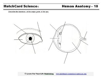
... Description Use these simple eye diagrams to help students learn about the human eye. Three differentiated worksheets are included: 1. Write the words using a word bank 2. Cut and paste the words 3. Write the words without a word bank Labels include: eyebrow, eyelid, eyelashes, pupil, iris, and sclera.
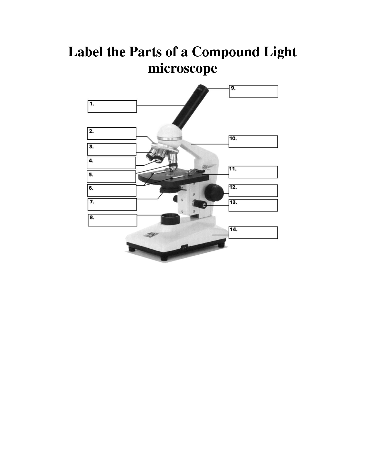
8 Best Images of Lens Diagram Worksheet - Microscope with Labeled Parts, Label Eye Parts ...
Quiz: Label The Parts Of The Eye - ProProfs Quiz How much did you get to understand about the human eye? Take up this quiz and find out! Questions and Answers. 1. A is pointing to what part of the eye? A. Cornea. B. Optic Nerve.
Human Eye Diagram
Anatomy of the Eye | Johns Hopkins Medicine Cornea. The clear, dome-shaped surface that covers the front of the eye. Iris. The colored part of the eye. The iris is partly responsible for regulating the amount of light permitted to enter the eye. Lens (also called crystalline lens). The transparent structure inside the eye that focuses light rays onto the retina. Lower eyelid.

Eye Diagram Without Labels | via Anatomy Pictures Gallery if… | Flickr
Human eye - Wikipedia The front part is also called the anterior segment of the eye. The eye is not shaped like a perfect sphere, rather it is a fused two-piece unit, composed of an anterior (front) segment and the posterior (back) segment. The anterior segment is made up of the cornea, iris and lens.

eye diagram - PurposeGames
Anatomy of the eye - Moorfields Eye Hospital Optic nerve: leaves the eye at the optic disc and transfers all the visual information to the brain. Sclera: the white part of the eye, a tough covering with which the cornea forms the external protective coat of the eye. Rod cells are one of the two types of light-sensitive cells in the retina of the eye. There are about 125 million rods ...

Activity Sheet 1: How the Eyes Work | Human eye diagram, Teaching biology, Human body activities
What Does the Eye Look Like? - Harvard Eye Associates Vitreous Gel: A thick, transparent liquid that fills the center of the eye. It is mostly water and gives the eye its form and shape. Our eyes are vital for seeing the world around us. Keep them healthy by maintaining regular vision exams. Contact Harvard Eye Associates at 949-951-2020 or harvardeye.com to schedule an appointment today.
8 Best Images of Lens Diagram Worksheet - Microscope with Labeled Parts, Label Eye Parts ...
PDF Parts of the Eye - National Institutes of Health Eye Diagram Handout Author: National Eye Health Education Program of the National Eye Institute, National Institutes of Health Subject: Handout illustrating parts of the eye Keywords: parts of the eye, eye diagram, vitreous gel, iris, cornea, pupil, lens, optic nerve, macula, retina Created Date: 12/16/2011 12:39:09 PM
Post a Comment for "41 parts of the eye without labels"